Serial Sections Of Chick Embryo


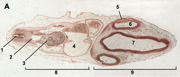
Contents • • • • • • • • Introduction Historically, an important way of teaching embryology is by direct observation of histological sections at different stages of development. Even today the best way to observe anatomical development and relationships is by direct observation in serial sections through the embryo and fetus. The sections below are from the original teaching set prepared at UNSW that were digitized and put online in 1996 as a teaching resource. The Stage 13 and 22 images are available in at least two formats, an unlabeled set (image 1 - 49) and a labeled set (image 50 - 98) in a rostro-caudal sequence (head to tail). Stage 22 embryo also has a set of selected images showing more detail of some organ and tissue development. In addition, there are now 3D animations based upon reconstruction of the embryos from these serial sections. 2011 - Selected slides are currently being rescanned at to show specific developmental details.
The Third Ear Lonsdale Pdf File. Author Comments Start here by looking through the early (week 4) embryo (stage 13) labeled images. Cooking Coke To Crack In A Spoon. It is not so important to identify every single feature, observe what structures are present and where they are in the middle of the embryonic period (week 4). Then if you like, look through the unlabeled images. What can you now recognise? Rocks Pebbles And Sand Rarest. Next look through the late (week 8) embryo (stage 22) labeled images as you did before. Then look through the same embryo stage selected labeled images.